Floor Of Carotid Triangle Diagram

Carotid Triangle Boundaries Contents Anatomyqa
Carotid Triangle Anatomy Of The Neck

Jaypeedigital Ebook Reader
Carotid Triangle Anatomy Kenhub

Carotid Triangle Boundaries Contents Anatomy Tutorial Youtube
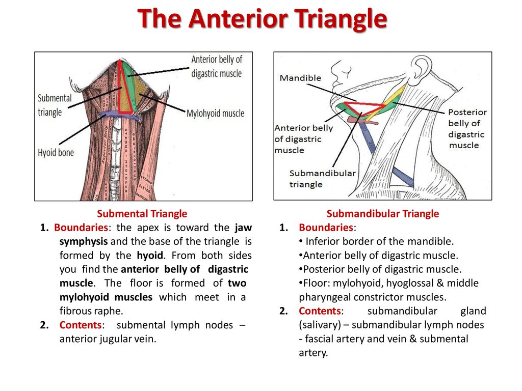
Anterior Triangle Of The Neck Subdivisions Teachmeanatomy

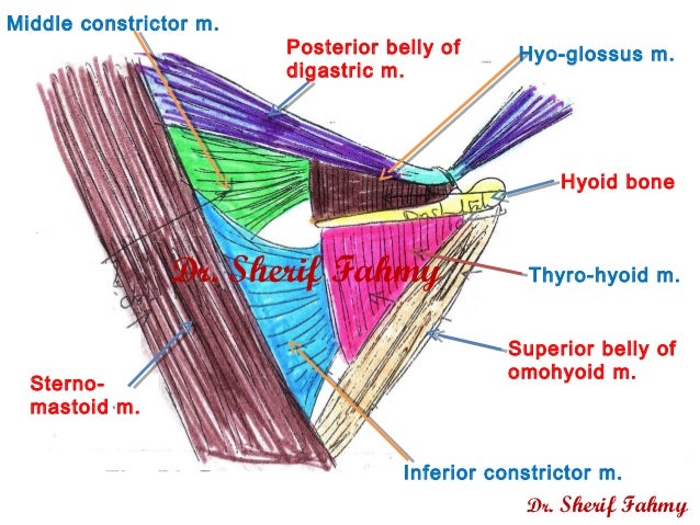
Hyoid bone thyro hyoid m.
Floor of carotid triangle diagram.

Jaypeedigital Ebook Reader

Carotid Triangle Of Neck 1 4 Youtube

Carotid Triangle 4 4 Content Diagram Youtube

Floor Of Femoral Triangle I P P Long Medicine Notes Human Anatomy And Physiology Anatomy

Case Based Learning Triangles Of Neck Region

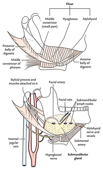
Contents Of Submandibular Triangle

Pdf Triangles Of The Neck A Review With Clinical Surgical Applications
Easy Notes On Anterior Triangle Of The Neck Learn In Just 3 Minutes Earth S Lab
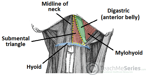
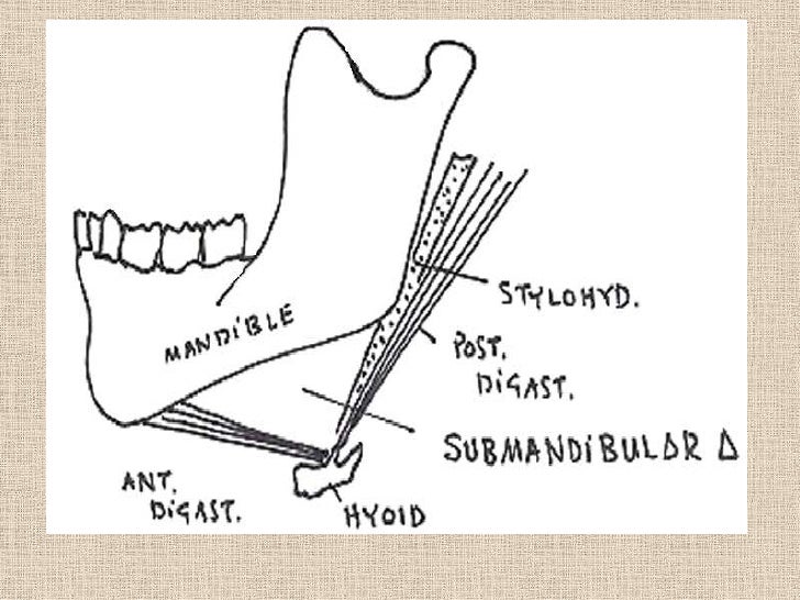
The Digastric Triangle
Surgical Anatomy Of Triangles Of Neck

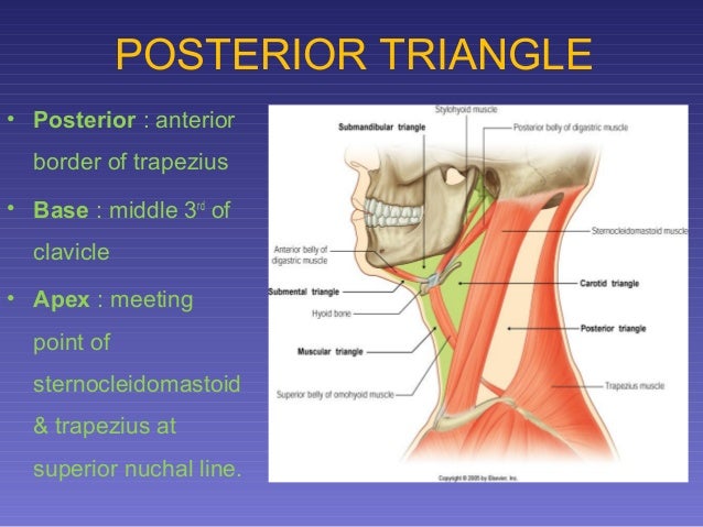
Posterior Triangle Of Neck Boundary Floor Youtube

Medicowesome Triangles Of The Neck Diagram And Mnemonic Mnemonics Medical Anatomy Anatomy And Physiology

Anterior Triangle Of The Neck

The Submandibular Gland Structure Vasculature Innervation Teachmeanatomy
10 Triangles Of Neck Tmj Applied Anatomy 1
Unit Iv Problem Iv Anatomy Ppt Download

Neck Anatomy Diagram Quizlet
Stylohyoid Origin Insertion Innervation And Action Kenhub
Https Encrypted Tbn0 Gstatic Com Images Q Tbn 3aand9gcr818buhdovpdkbniuybyw8tdoxzc6o1uudqgg73 A2aeyg8ntx Usqp Cau

Instant Anatomy Head And Neck Nerves Cranial Xi Spinal Accessory In Posterior Triangle Neck Muscle Anatomy Head And Neck Human Anatomy And Physiology

Neck Triangles Diagram Quizlet
Https Judoctor2011 Files Wordpress Com 2013 02 Neck Part 2 Pdf

Anal Triangle Wikipedia
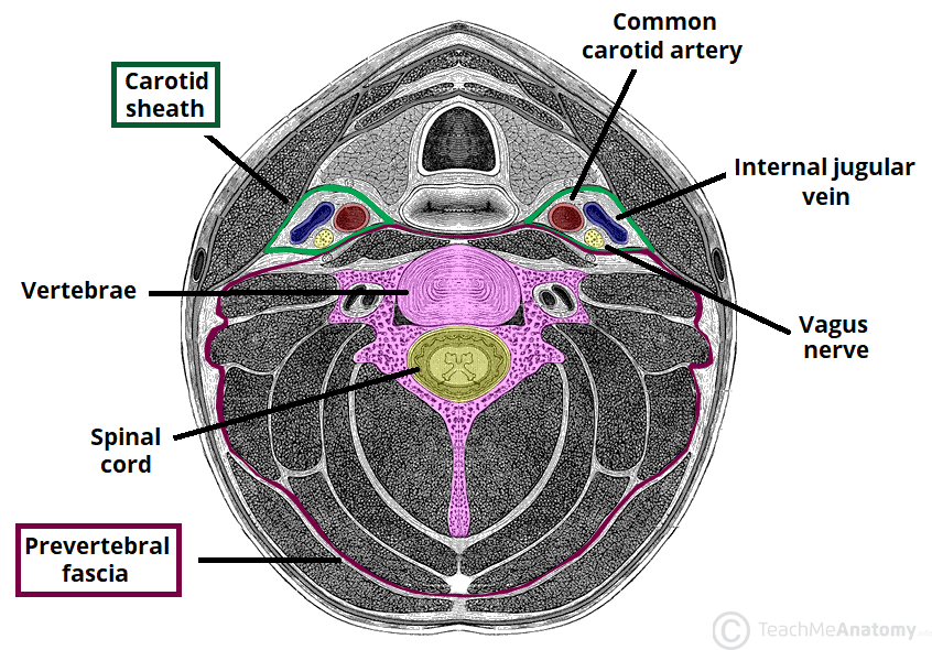
Fascial Layers Deep Superficial Teachmeanatomy
Source : pinterest.com
