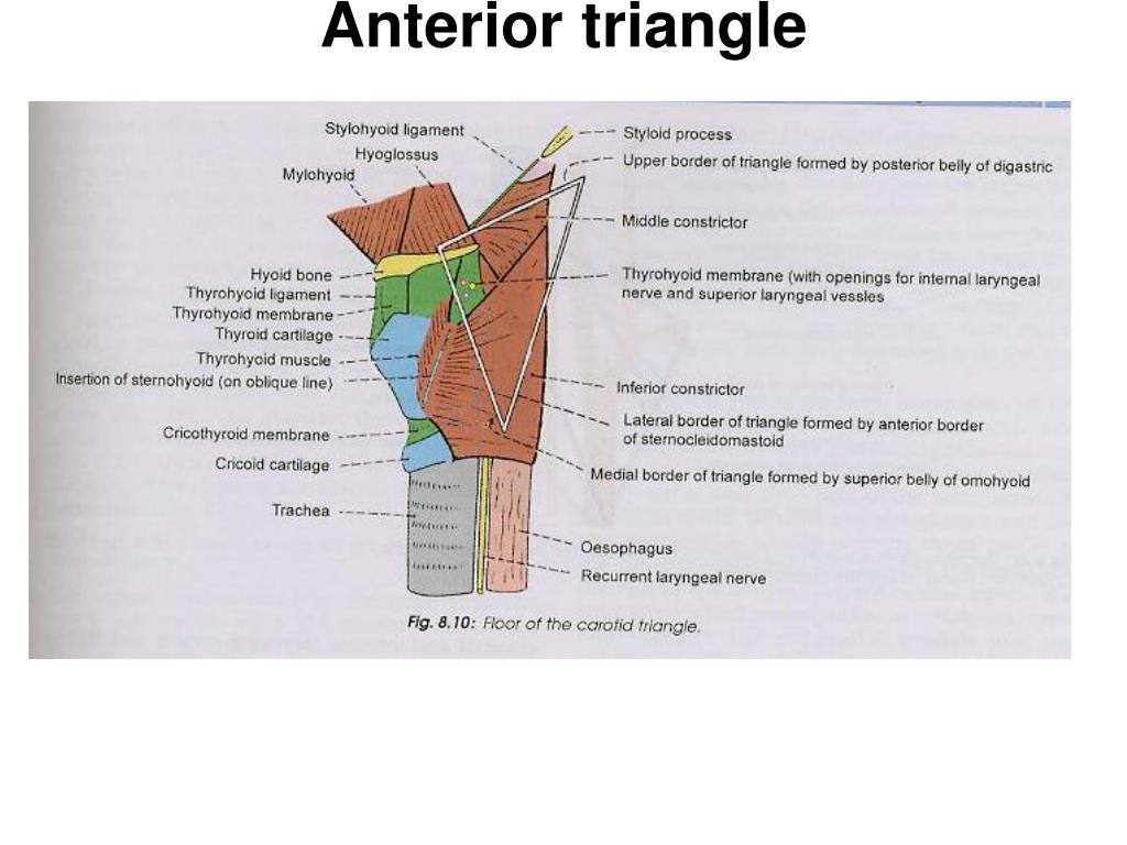
Floor Of Carotid Triangle Is Formed By

Triangles Of The Neck Diagram And Mnemonic Mnemonics Medical Anatomy Anatomy And Physiology
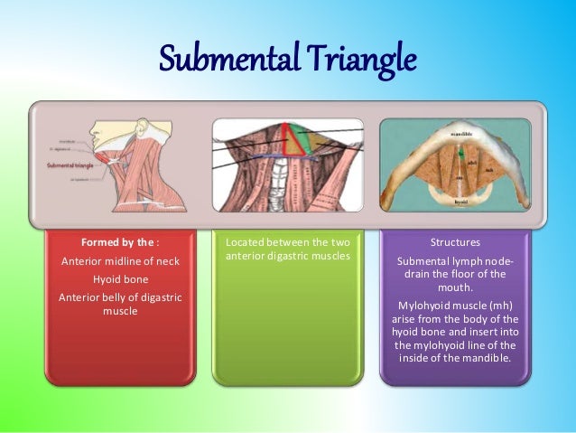
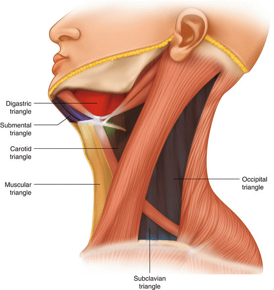
Triangles Of The Neck
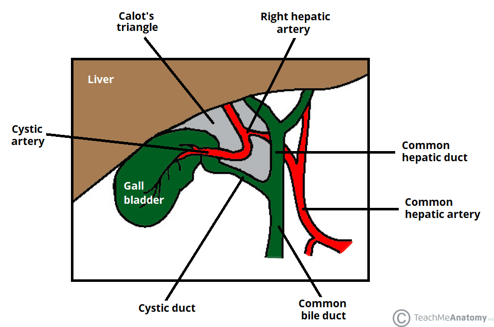
Calot S Triangle Borders Contents Cholecystectomy Teachmeanatomy
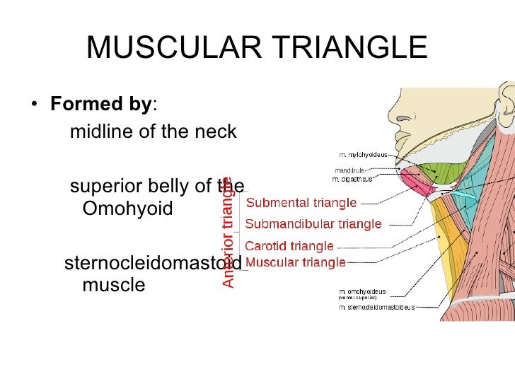
Triangles Of The Neck Ppt Year 1
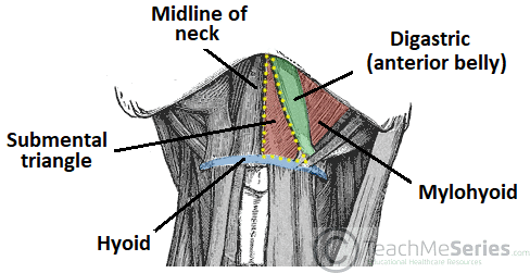
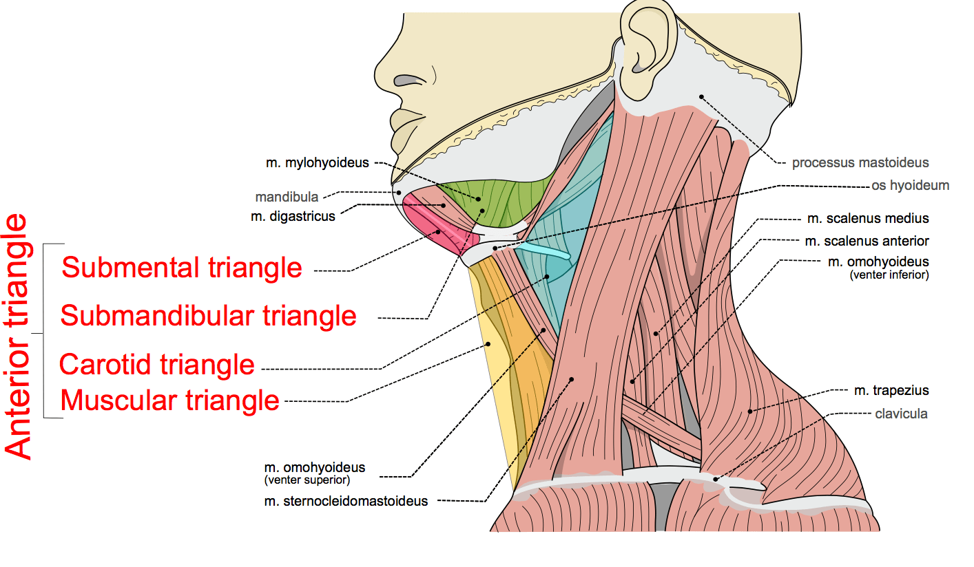
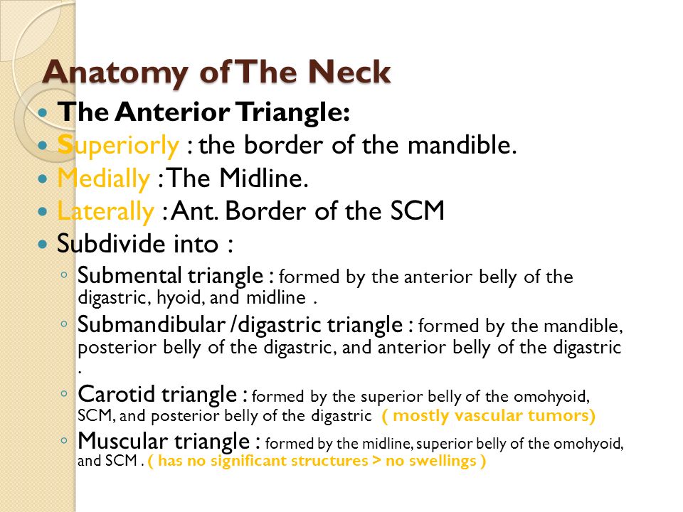
Anterior Triangle Of The Neck Subdivisions Teachmeanatomy

Carotid Triangle Anatomy Kenhub
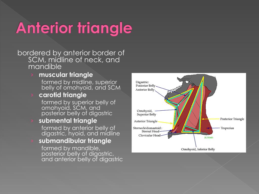
The triangles of the neck are important because of their contents as they house all the neck structures.
Floor of carotid triangle is formed by.

Occipital Triangle Wikipedia

Triangles Of The Neck Anatomy Borders And Contents Kenhub

Neck 1 Superficial Neck Posterior Triangle Cervical Viscera Flashcards Quizlet
Triangles Of The Neck Part 1 The Anterior Triangle Medical Exam Prep

12 Neck Anatomy Trebloc Flashcards Quizlet

Vagus X Cranial Nerves Cranial Nerves Nerve Vagus Nerve

Anterior Triangle Of The Neck

Carotid Triangle Boundaries Contents Anatomy Tutorial Youtube

Mylohyoid And Related Structures Suspended From The Body Of The Mandible Is A Thin Sheet Of Muscle Fo Glossopharyngeal Nerve Sensory Nerves Hypoglossal Nerve

Inguinal Canal Medical Anatomy Pelvis Anatomy Human Anatomy And Physiology

Lumbar Triangle 02 Petit S Triangle Note Floor Is Formed By Internal Oblique Muscle Human Body Anatomy Body Anatomy Medical Anatomy

Posterior Triangle Of The Neck Everything You Need To Know Dr Nabil Ebraheim Youtube
Swellings Of The Neck Springerlink
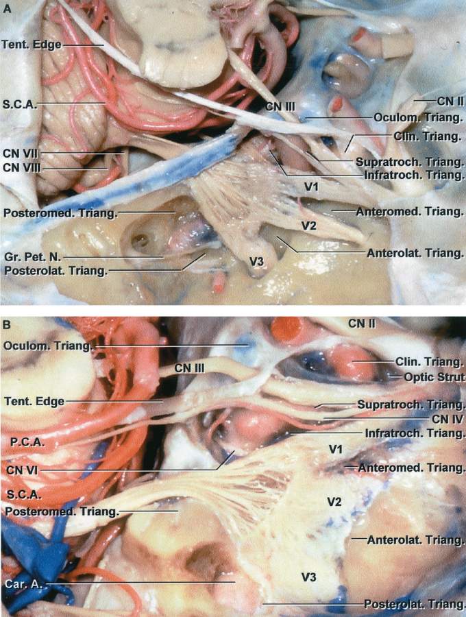
Triangles In The Region Of The Cavernous Sinus And Middle Fossa Formed By The Convergence And Divergence Of The Cranial Nerves Neuroanatomy The Neurosurgical Atlas By Aaron Cohen Gadol M D
Ppt Neck Mass Powerpoint Presentation Free Download Id 6059901
Https Ispub Com Ijha 2 1 8323

Vascular Cannulation Anesthesia Key

Triangles Of Neck Made Easy Youtube
1
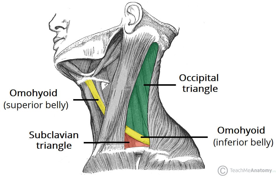
Posterior Triangle Of The Neck Subdivisions Teachmeanatomy
Lateral Neck Masses Prof Alam Ppt Video Online Download
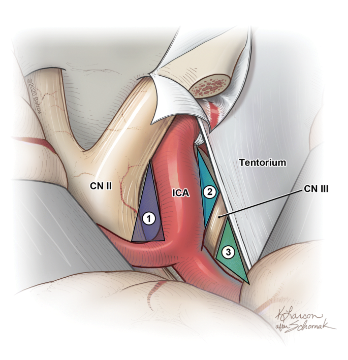
Tailoring The Surgical Corridor To The Basilar Apex In The Pretemporal Transcavernous Approach Morphometric Analyses Of Different Neurovascular Mobilization Maneuvers Springerlink
Ppt Anterior Triangle Powerpoint Presentation Free Download Id 5436377

Lumbar Triangle Wikipedia
Source : pinterest.com
