Floor Of Left Maxillary Sinus
Panoramic Radiograph Of The Case Opacification Of Left Maxillary Download Scientific Diagram
Benign Maxillary Sinus Masses Ento Key

Cbct Images Of Left Maxillary Sinus In Sagittal View Depicting Relation Download Scientific Diagram

Thick Inspissated Mucous Covering The Left Maxillary Sinus Floor Download Scientific Diagram
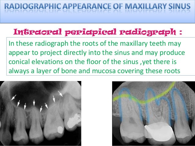
Radiology Of Maxillary Sinus

Isolated Unilateral Upper Alveolar Numbness In Silent Sinus Syndrome Bmj Case Reports

Among the diseases are sinusitis frontal sinusitis etmoiditois and.
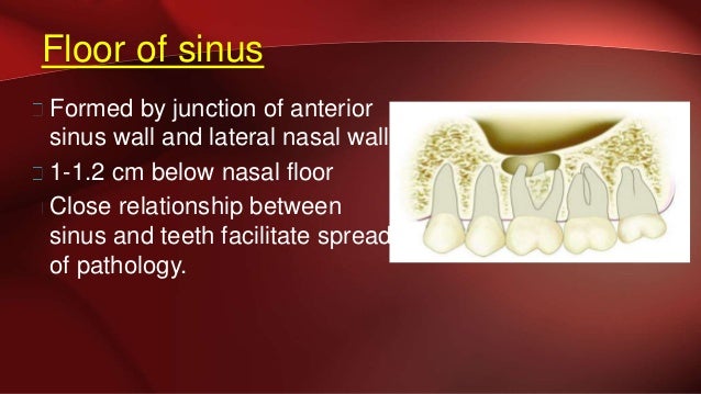
Floor of left maxillary sinus.

Postoperative Stage 2 Panoramic Radiograph Of Left Maxillary First Download Scientific Diagram

Cranio Facial Ct Scan Coronal Sections Left Maxillary Sinus With Download Scientific Diagram

Pns View Showing Destruction Of Floor Of Maxillary Antrum On Left Side Download Scientific Diagram

Intraoperative Endoscopic Views Of The Left Maxillary Sinus Of An Orbital Floor Fracture Through The Antral Window A The Orbital Floor Was Fractured And Periorbital Soft Tissue Was Herniated Into The Maxillary

Emergency Department Computed Tomography Showing A Thickened Inflamed Download Scientific Diagram
Com Sept 2019 Uw School Of Dentistry
Maxillary Sinus Certified Fixed Orthodontic Courses By Indian Dental

Maximum Height Of The Medial Maxillary Sinus Roof Relative To The Download Scientific Diagram

Evaluation Of Haller Cell On Cbct And Its Association With Maxillary Sinus Pathologies Kamdi P Nimma V Ramchandani A Ramaswami E Gogri A Umarji H J Indian Acad Oral Med Radiol

Radiograph Of The Paranasal Sinuses Water S View Reveals A Small Left Download Scientific Diagram
Figure 5 The Prevalence Of Concha Bullosa And Nasal Septal Deviation And Their Relationship To Maxillary Sinusitis By Volumetric Tomography

Figure 6 From Odontogenic Maxillary Sinusitis Need For Multidisciplinary Approach A Review Semantic Scholar

Pdf Case Report Retained Gutta Percha As A Cause For Persistent Maxillary Sinusitis And Pain

Nasal Cavity Paranasal Sinuses Radiology Key

Jordi Journal Of Oral Diagnosis

Pdf Silent Sinus Syndrome Report Of A Case

A And B Coronal And Sagittal Ct Showing Superior Bowing Of The Right Download Scientific Diagram
Odontogenic Maxillary Sinusopathies A Radiological Classification Springerlink
Https Encrypted Tbn0 Gstatic Com Images Q Tbn 3aand9gct55cwhybwpk2x296c66clurkwjubxkriq3d9o40f5ykmielddm Usqp Cau

The Cbct Showed Veiling Of The Left Maxillary Sinus Soil Discontinuity Download Scientific Diagram

Assessment Of Prevalence Of Anatomical Variations And Pathosis Of The Maxillary Sinuses Using Cone Beam Computed Tomography In A Sample Of The Population Of Saudi Arabia Alzain L Alzain S Badr F Ashy

Pdf Panoramic Radiographs Are Of Limited Value In The Evaluation Of Maxillary Sinus Disease
Extensive Complex Odontoma In The Maxillary Sinus Pushing 3rd Molar Near The Orbital Floor Causing Transient Diplopia And Chronic Sinusitis A Rare Presentation And Surgical Management Springerlink
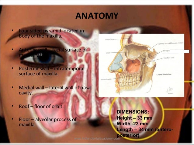
Maxillary Sinus
Source : pinterest.com
