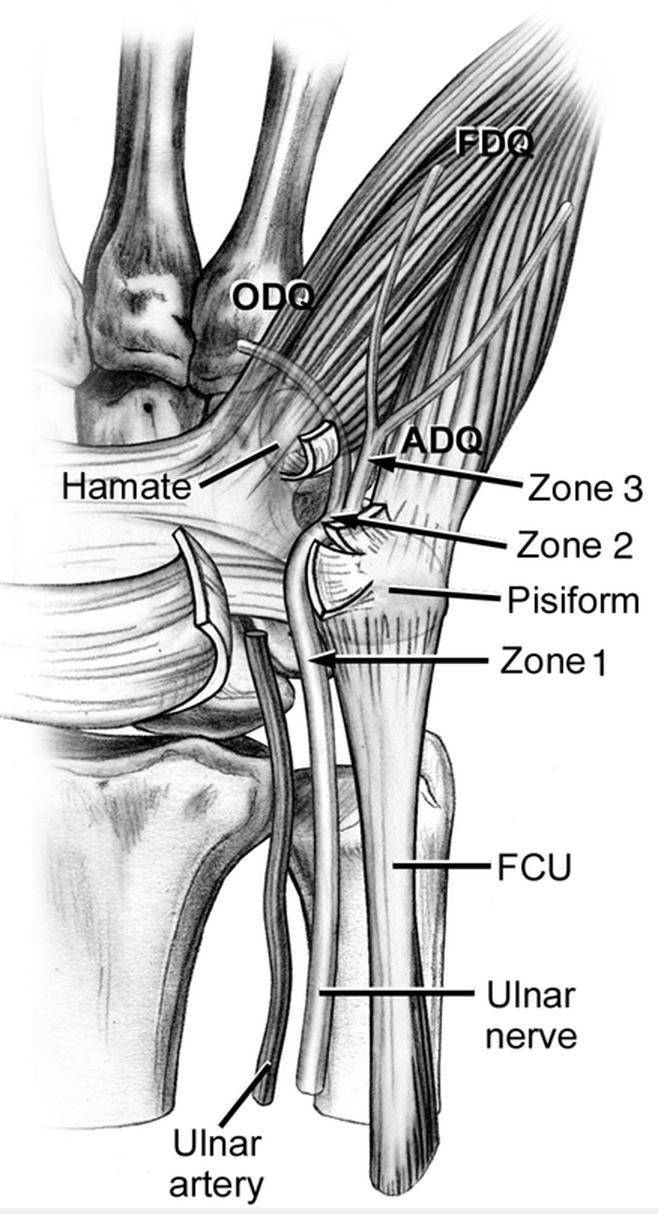
Floor Of The Distal Ulnar Tunnel
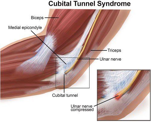
Physical Therapist S Guide To Cubital Tunnel Syndrome

Ulnar Nerve Entrapment An Overview Sciencedirect Topics

Cubital Tunnel Syndrome Sciencedirect

47 Ulnar Tunnel Syndrome Guyon S Canal Syndrome Radiology Key
Ulnar Neuropathy Causes Symptoms Diagnosis Treatment Exercises

Cubital Tunnel Syndrome Ulnar Nerve Entrapment Kansas City Bone Joint Clinic
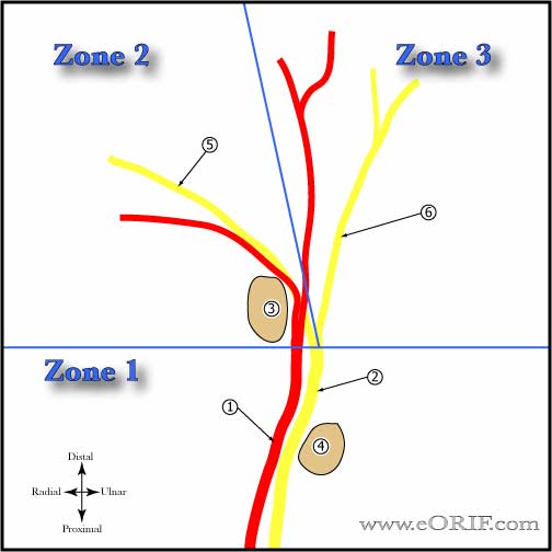
1 the triangular shaped loge contained the ulnar nerve and artery with accompanying.
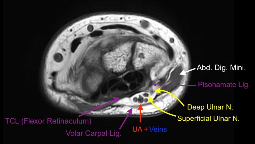
Floor of the distal ulnar tunnel.
Ulnar Tunnel Syndrome Features And Treatment Bone And Spine
Ulnar Tunnel Syndrome Springerlink

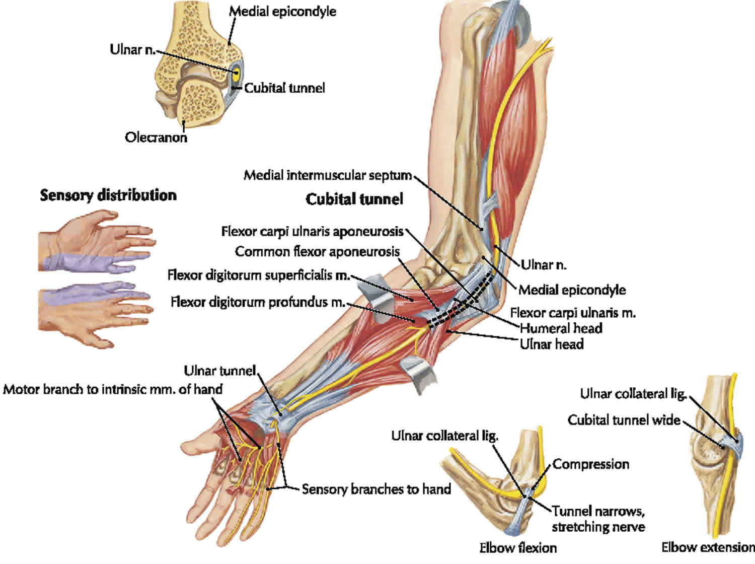
Ovid Lippincott Williams Wilkins Atlas Of Anatomy Ulnar Nerve Entrapment Ulnar Nerve Nerve Entrapment

Ovid Lippincott Williams Wilkins Atlas Of Anatomy Ulnar Nerve Entrapment Ulnar Nerve Nerve Entrapment

Incision For The Release Of Cubital Tunnel Syndrome Download Scientific Diagram
Bilateral Anconeus Epitrochliaris Causing Cubital Tunnel Syndrome A Case Report

Ulnar Nerve Compression Living Handbooks

Pdf Diagnosis And Treatment Of Work Related Ulnar Neuropathy At The Elbow

That Funny Bone Erik Dalton Blog

Cubital Tunnel An Overview Sciencedirect Topics
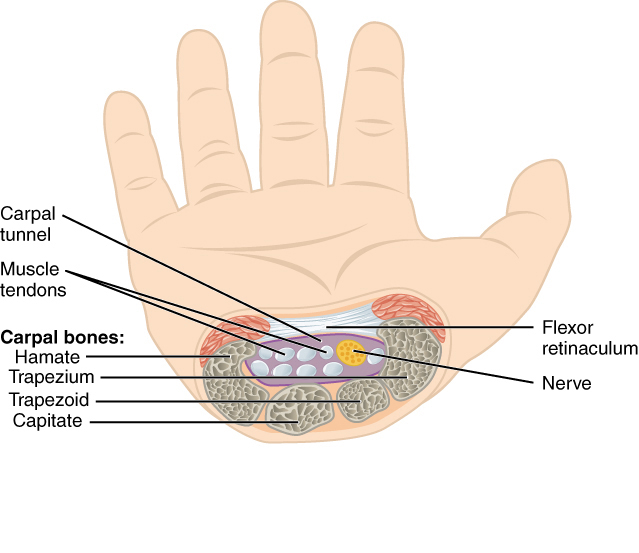
Carpal Tunnel Wikipedia

Ulnar Nerve Msk Medbullets Step 1

View Of The Wrist Showing The Flexor Retinaculum At The Wrist And The Carpal Tunnel Where The Median Ne Hand Therapy Carpal Tunnel Human Anatomy And Physiology

The Cubital Tunnel Syndrome A Case Report And Discussion Sciencedirect

Bijopaul Cubital Tunnel Syndrome
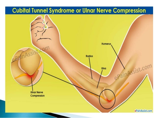
Cubital Tunnel Syndrome

Pin On Joint Pain

Bones Of The Hand Human Anatomy And Physiology Anatomy And Physiology Hand Anatomy
1
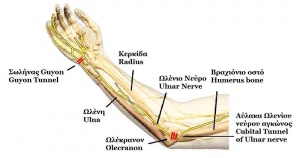
Ulnar Nerve Entrapment Physiopedia
Guyon S Canal Radsource

Carpal Tunnel Surgery Wikipedia
Ulnar Tunnel Syndrome S64 00xa 354 2 Eorif

Picture Test In Anatomy Wrist And Hand 1 Anatomy Carpal Tunnel Syndrome Carpal Tunnel
Source : pinterest.com
